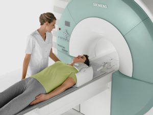
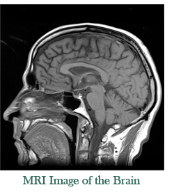
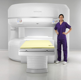
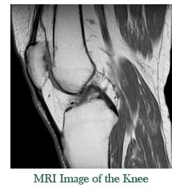
Magnetic resonance imaging (MRI) is a non-invasive way of acquiring high quality images of internal body structures. MRI uses radio waves passed through a powerful magnetic field to produce clear and detailed pictures of the body, providing information that cannot be otherwise obtained from an x-ray, ultrasound, or computed tomography (CT) scan. Some MRI studies include an injection of gadolinium contrast media (“dye”) to gather additional information. MRI exam length varies from 20 minutes to an hour or more, depending on the specific exam and types of images needed for an accurate diagnosis. MRIs can be performed on any part of the body, but are most frequently performed on the brain, spine, abdomen, pelvis, breast, and large joints like the knee and shoulder.
We perform all routine MRI imaging studies and advanced MRI exams including MRAs and breast imaging (including MRI-Guided Breast Biopsies).
We offer traditional MRI exams in Fort Collins in our 1.5T Siemens Aera big bore scanners, and even high-strength 1.2T open MRI exams at our Loveland/Johnstown location.
To learn more about MRI, please visit www.radiologyinfo.com
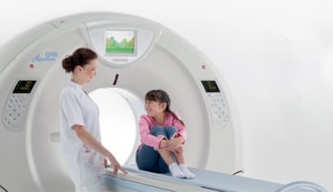
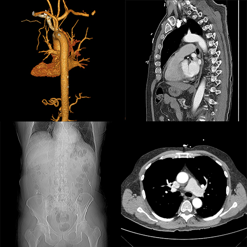
Computed Tomography (CT), sometimes called CAT scan, uses special x-ray equipment to gather image data from different angles around the body and then uses computer processing of the information to show a cross-section of body tissues and organs. Some CT exams may require you to drink a special dye and/or to have an injection of contrast media (“dye”). Most CT exams only take a few minutes on the exam table. Learn More
We perform all routine CT studies and advanced studies such as CTA, Calcium Scoring, CT Low-Dose Lung Screening, TAVR, and Virtual Colonoscopy.
To learn more about CT, please visit www.radiologyinfo.com

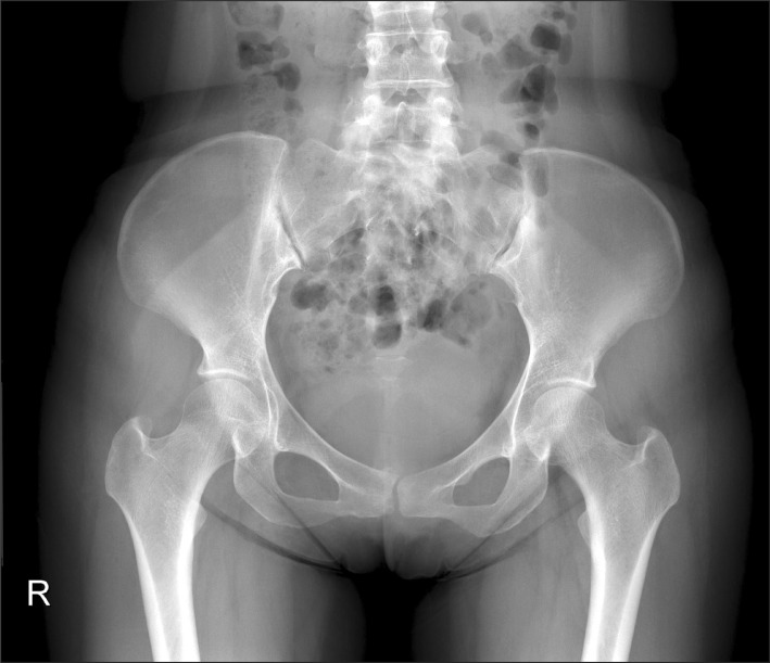
An x-ray is a painless medical test that helps physicians diagnose and treat medical conditions. Radiography involves exposing a part of the body to a small dose of radiation to produce pictures of the inside of the body. X-rays are the oldest and most frequently used form of medical imaging. We also perform fluoroscopy studies to gather live images of internal organs including esophagrams, upper GI studies, barium enema studies, IVPs, arthrograms, and pain injections.
To learn more about X-Ray, please visit www.radiologyinfo.com
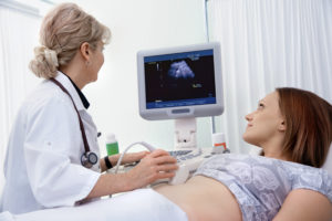
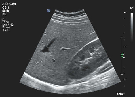
Ultrasound imaging, also called ultrasound scanning or sonography, involves exposing part of the body to high-frequency sound waves to produce pictures of the inside of the body. Ultrasound exams do not use ionizing radiation (x-ray). Because ultrasound images are captured in real-time, they can show the structure and movement of the body’s internal organs, as well as blood flowing through blood vessels.
To learn more about Ultrasound, please visit www.radiologyinfo.com
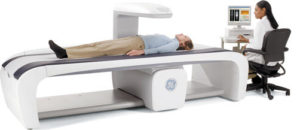
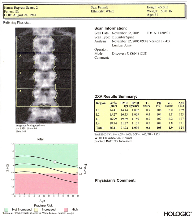
The DEXA bone density scan uses advanced X-ray technology to examine bones, determining how dense they are and how much bone mass has been lost. There are several tests and methods that can be used to diagnose bone loss, but the DEXA bone density scan is considered the gold standard. A DEXA bone density scan is generally recommended for people over age 65 and those who have risk factors for osteoporosis. The scan can tell you the status of your bone health and help your doctor determine what steps you need to take should the results indicate bone loss and osteoporosis risk.
To learn more about bone density scans, please visit www.radiologyinfo.com